Showing posts with label carcinoma. Show all posts

Tuesday, July 27, 2021

Ductal Vs Lobular Carcinoma

Lobular may be a problem in pagetoid or complete involvement of ducts by LCIS in solid low grade DCIS or in lobular involvement by DCIS cells cancerization of lobules DCIS LCIS. Ductal carcinoma can remain within the ducts as a noninvasive cancer ductal carcinoma in situ or it can break out of the ducts invasive ductal carcinoma.

Lobular Carcinoma In Situ Wikipedia

Invasive carcinoma can be ductal or lobular but lobular type is much more common Ipsilateral invasive lobular carcinoma is three times more common than contralateral invasive lobular carcinoma LCIS is no longer classified as Tis because it is considered a risk factor not a malignancy Amin.

Ductal vs lobular carcinoma. ObjectiveInvasive lobular carcinoma ILC and invasive ductal carcinoma IDC account for most breast cancers. Lobular carcinoma begins in the lobes or lobules glands that make breast milk. The word invasive means.

An analysis of the largest recorded cohort of patients with invasive lobular breast cancer ILC demonstrates that outcomes are significantly worse when compared with invasive ductal breast cancer IDC highlighting a significant need for more research and clinical trials on patients with ILC. P 0001 and after matching for stage T1N0 98 vs. Classic variant of invasive lobular carcinoma of any grade has better prognosis than nonclassic variants overall Cancer 20081131511 Am J Surg Pathol 19901412 May have similar long term prognosis as infiltrating ductal carcinoma Breast Cancer Res Treat 2009117211 but see J Clin Oncol 2008263006 lobular has better survival at 6 years but worse survival at 10 years.

The 5-year disease-specific survival DSS was significantly better for patients with ILC than for those with IDC before 90 vs. This study aimed to compare nonmetastatic ILC to IDC in terms of survival and prognostic factors for ILCMethodsThis retrospective cohort study used data from the Surveillance Epidemiology. Lobular tumors are often slower growing than ductal tumors are more often estrogen and progesterone receptor positive 3 4 have lower vascular endothelial growth factor expression 5 and more frequently have loss of E-cadherin 6 7 8.

From the clinical standpoint data suggest that ILC derives a distinct benefit from systemic therapy compared to IDC. Invasive lobular carcinoma ILC is the second most common histologic subtype of breast cancer BC. P 0001 lymph node positive 368 vs.

This type of cancer is called invasive ductal carcinoma IDC. Lobular tumors have a tendency to metastasize to more axillary lymph nodes than ductal tumors of the same grade or size. P 0001 and T3N0 92 vs.

We compared biological and clinical characteristics of HER2-positive ILC versus HER2-positive infiltrating ductal carcinoma IDC. Ductal carcinoma in situ DCIS is the most common type of non-invasive breast malignancy and currently comprises around 20 of all breast cancers diagnosed. Most people with breast cancer have the disease in their ducts which are the structures that carry milk.

However the overall survival OS differences between ILC and IDC remain controversial. It begins in the lobules. It is a malignancy of the ductal tissue of the breast that is contained within the basement membrane Fig.

Lobular breast cancer tends to be diagnosed at a later stage and larger tumor size than ductal breast cancer. We used a national registry to compare outcomes of patients with stage-matched ILC and IDC. 2 yet 20-30 of cases who do not receive treatment will develop invasive disease.

The lobules are connected to the ducts which carry breast milk to the nipple. Infiltrating lobular carcinoma ILC represents about 10 of breast cancer and rarely shows overexpression of human epidermal growth factor receptor 2 HER2. When it breaks out of the lobules its considered invasive lobular carcinoma.

Ductal Carcinoma in Situ. Does delayed diagnosis of ILC affect survival. When compared with IDC ILC was more likely to be 2 cm 431 vs.

P 0001 T2N0 94 vs. ILC differs from invasive ductal carcinoma IDC in its clinicopathological characteristics and responsiveness to systemic therapy. It was found that patients with.

Understanding the differences between these two specific types is crucial in the proper prevention and treatment of the disease. Lobular carcinoma in situ LCIS. Lobular carcinoma starts in the lobules of the breast where breast milk is produced.

AJCC Cancer Staging Manual 8th Edition 2016. P 0001 and ER positive 931 vs. Ductal and lobular carcinomas are deadly because of their aggressive nature.

This study compared mixed invasive ductal and lobular carcinoma IDC-L with invasive lobular carcinomas ILCs to assess the overall prognosis the prognostic role of histologic grade and response to systemic therapy. Invasive lobular breast cancer ILC is less common than invasive ductal breast cancer IDC more difficult to detect mammographically and usually diagnosed at a later stage. Chemotherapy or surgery are usually the first options your doctor will recommend for infiltrating ductal or lobular carcinoma.

Sunday, September 8, 2019

Metastatic Lung Carcinoma

By the time lung cancer is metastatic it is in a late stage and can be challenging. This patient had renal cell carcinoma which is often said to give rise to canon-ball metastases in the lungs - a descriptive term referring to their appearance -.

Metastatic Lung Carcinoma And Other Metastatic Neoplasms A A Focal Download Scientific Diagram

Metastatic cancer has the same name as the primary cancer.

Metastatic lung carcinoma. It is treated as stage IV breast cancer not as lung cancer. The spread of cancer is called metastasis. Sometimes when people are diagnosed with metastatic cancer.

Metastatic lung cancer is cancer that starts somewhere else in the body and spreads to the lungs. Lung cancer might be classified as metastatic upon initial diagnosis or later on following treatment. Ad Welcome to SIE Medical where we take the time to treat you as an Individual.

They suggest a metastatic mode of spread involving the renal lymphatic drainages and specific biologic characteristics acquired by. Learn about metastatic lung cancer which occurs when lung cancer cells break away from a tumor and travel to other parts of your body through the blood or lymph system. Adenocarcinoma metastases rather than displace or destroy adjacent lung parenchyma may grow in a lepidic fashion spread along alveolar walls resulting in pneumonia-like consolidation.

Metastatic cancer to the lung. Metastases to lung - Renal cell carcinoma. Among all solid tumors small cell lung cancer SCLC has remarkable metastatic proclivity with a majority of patients diagnosed with metastatic disease.

In about 70 of cases the cancer has already spread to other areas of the body by the time of. Renal cell carcinoma lung metastases surgery. Air bronchograms may also be visible 1.

About 85 percent of lung cancers are non-small cell. Ad Welcome to SIE Medical where we take the time to treat you as an Individual. Metastatic lung cancer is the most advanced lung cancer stage where the disease has spread metastasized to distant organs.

Treatment for lung metastases is usually based on the main type of cancer primary site the person has. For example breast cancer that spreads to the lung is called metastatic breast cancer not lung cancer. It is sometimes referred to as stage 4 cancer.

Cavitation is present in 4 of cases 1. Size of LM and lymph node involvement are important prognostic factors. Our understanding of SCLC metastasis has been hampered for many years by the paucity of material from primary tumors and metastases as well as the lack of faithful pre-clinical models.

Lung cancer can be metastatic at the time of diagnosis or following treatment. It can occur with any type of lung cancer. Treatment may include chemotherapy immunotherapy or radiation therapy or a combination of these.

What this means is that cancer cells can separate themselves from a tumor and travel through the blood or lymph system to other areas in the body. There are two main types of lung cancer. Small cell lung cancer accounts for about 1015 of all lung cancer cases.

Pathologic findings and prognostic factors. Metastatic lung cancer is lung cancer that has started to spread. Metastatic lung cancer is also called stage 4 lung cancer.

Metastatic tumors in the lungs are malignancies cancers that developed at other sites and spread via the blood stream to the lungs. Surgery may be an option if there are a small number of lung metastases and there are no metastases in other parts of the body. Metastatic lung cancer begins in the lungs and spreads to other areas.

This image shows numerous lung nodules of varied sizes scattered throughout both lungs.

Tuesday, January 1, 2019

Afp Hepatocellular Carcinoma

Some guidelines have advised against the use of AFP in the diagnosis of HCC. If caught early it can sometimes be.

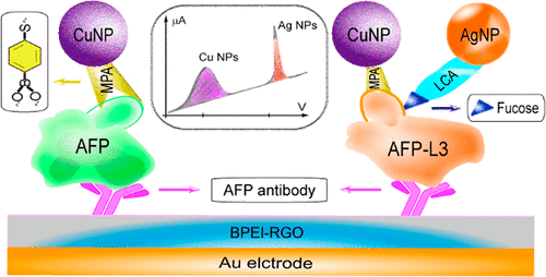
Electrochemical Assay Of The Alpha Fetoprotein L3 Isoform Ratio To Improve The Diagnostic Accuracy Of Hepatocellular Carcinoma Analytical Chemistry X Mol

In addition AFP is also an indicator of HCC risks mostly in patients with cirrhosis and HCVHBV infections.

Afp hepatocellular carcinoma. It is a glycoprotein produced by the fetal liver and yolk sac during pregnancy. Among the study population 96. AFP level is elevated in 60 of patients with HCC typically those with the most advanced disease.

It is concluded that AFP is a significant markers for Hepatocellular carcinoma helpful in assessing problems in management of HCC and monitoring treatment regiments. AFP is serum glycoprotein produced by fetal yolk sac and fetal liver tissue and it drops rapidly after the birth under 10ngmL. Symptoms and signs are usually nonspecific.

Therefore AFP is the most used marker for detecting HCC. Its different from secondary liver cancers which have spread to the liver from other organs. Hepatocellular carcinoma HCC is the fourth leading cause of cancer-related death worldwide.

Hepatocellular carcinoma usually occurs in patients with cirrhosis and is common in areas where infection with hepatitis B and C viruses is prevalent. As a diagnostic criteria of HCC the exact threshold of Alpha-fetoprotein AFP is controversial. Alphafetoprotein AFP is the most widely accepted serum biomarker used in the management of hepatocellular carcinoma HCC.

Hepatocellular carcinoma HCC has become a pressing health problem facing the world today due to its high morbidity high mortality and late discovery. The sensitivity ranges from. Hepatocellular carcinoma is responsible for over 12000 deaths per year in the United States where the incidence of the disease is approximately 25 per 100000 population.

A median follow-up of 654IQR 13-114 months recurrence occurred in 648 70108 patients. Mild elevations may occur in patients with chronic hepatitis without HCC. Diagnosis is based on alpha-fetoprotein AFP levels imaging tests and sometimes liver biopsy.

Therefore AFP levels are also frequently elevated in chronic active hepatitis. AFP can be normal in significant number of patients with HCC. The first serologic assay for detection and clinical followup of patients with hepatocellular carcinoma was alpha-fetoprotein AFP which has been the standard tumor biomarker for HCC for many years.

The role of serum α-fetoprotein AFP measurements in the diagnosis of hepatocellular carcinoma HCC remains controversial. Hepatocellular carcinoma is a tumor of the liver. One hundred and eight patients fulfilled the inclusions criteria.

The regulation and pathophysiology of AFP informs the basis for its clinical relevance in the context of HCC. 16 rows Patients can be screened for hepatocellular carcinoma using AFP. Alpha-fetoprotein AFP is the standard serum tumor marker utilized in the evaluation of suspected hepatocellular carcinoma.

Although the current application of immunotherapy for HCC is still on trial immunotherapy has achieved certain effect in the treatment of liver cancer. AFP plays an important role in the development of hepatocellular carcinoma which can serve as a target for immunotherapy of HCC. Hepatocellular carcinoma HCC is expected to become a leading cause for liver transplantation LT following curative treatment for hepatitis C and after more widespread acceptance of the practice of tumour down-staging for patients originally considered beyond LT criteria.

1 Many clinical factors are important in the prognosis of patients with HCC including disease stage. Hepatocellular carcinoma is a cancer that starts in your liver. HCC cells produce AFP and the serum level of AFP is usually increased in patients with HCC.

It is one of the most common malignancies in adults and is more common in men than women 2-41 and blacks than whites. The secondary end points were overall survival OS and disease-free RESULTS. AFP levels may be elevated because of production by the tumor or by regenerating hepatocytes.

Side Effects Of Drinking Alcohol

Many people who arent regular alcohol drinkers like to imbibe on special holidays or occasions but the next morning they invariably kick th...